Our Services

Endoscopic Department

The Endoscopy Department of EIMC is located in Athena Polyclinic (part of EIMC HealthCare Clinics), in Larnaca, and is fully equipped to perform endoscopic examinations with absolute safety. The Endoscopic Department performs diagnostic and therapeutic endoscopic procedures by the pulmonologists and gastroenterologists of EIMC with the assistance of our excellent nursing staff.  All endoscopic procedures are performed in the best possible way, with the sole aim of the well-being of our patients!

Endoscopy

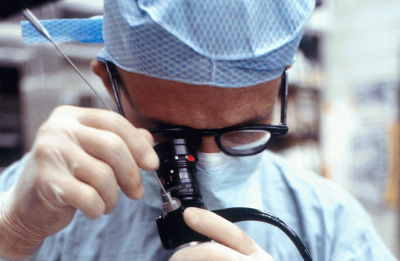
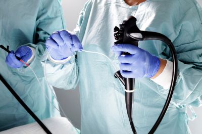
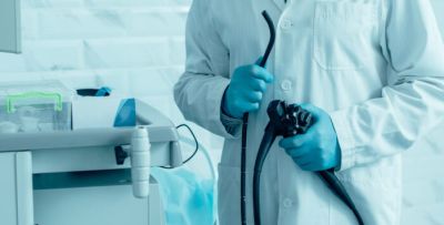
Endoscopy is the process which enables the doctor to look inside the human body with the use of an endoscope, a slender and tubular instrument with an illuminated, guided video camera. Since this technique allows the doctor to insert the endoscope directly into the area of interest, the doctor can perform procedures like visual diagnosis, biopsies, and removal of polyps if needed. There are various kinds of endoscopies for different body areas, such as bronchoscopy, gastroscopy, and colonoscopy.

Osteopathy, Medical Acupuncture

Osteopathy involves clinical care of the neuro-musculoskeletal system, which is made up of the bones, muscles, nerves and other tissues that support your body and control its movements.
Osteopaths use your individual medical history and environmental factors to understand the root cause of your pain to develop a personalised plan to help you manage your pain and move better.
Osteopathic treatment may involve any combination of Manual Therapy (manipulation, mobilisation, or neuromuscular techniques, soft tissue massage), Therapeutic Needling Techniques and Clinical Exercise Program. The treatment is tailored to each individual and is encouraged through advice on posture, lifestyle and aid to stress-related problems. This clinical advice aims to help patients understand the nature of their problems so that they can return to their everyday activities as soon as possible.

Bronchoscopy

Bronchoscopy is the endoscopic method used by doctors to have a look at the inside of the patient’s airways for diagnostic and therapeutic purposes. The doctor inserts the bronchoscope into the patient’s airways through the nose or mouth (and rarely through a tracheostomy) to operate with anomalies such as tumors, bleeding, foreign bodies, or inflammation. The bronchoscope is an advanced flexible device that contains lighting with video equipment and enables doctors to make a more precise examination and allows them to collect specimens from inside the lungs.

When does a patient need a bronchoscopy?

The main reason is a continuing cough, illness or the doctor identified something unusual on an X-ray or other kind of test. Of course, it can be used for other anomalies.

Requirement before and after bronchoscopy:

You may need to stop taking some anticoagulants a few days before the test, but it is best to ask your doctor. Eat a light dinner the night before and NEVER eat or drink anything on the morning of the test. You will also need to remove any dentures, hearing aids, contact lenses, or eyeglasses you may be wearing. At the end of the test, you will eat or drink three hours later. It is also essential to be accompanied by a relative or a friend since you will not drive after the examination.

Gastroscopy

Gastroscopy — also known as upper endoscopy — involves using a thin flexible tube (endoscope) to examine the upper digestive tract. The gastroenterologist, assisted by a nurse, inserts the endoscope into the mouth and travels down the food pipe (esophagus), then into the stomach and first part of the small intestine (duodenum), to view these areas. Gastroscopy usually lasts about 15 minutes.

When does a patient need a gastroscopy?

A gastroscopy helps your doctor to determine what’s causing digestive signs and symptoms like nausea, vomiting, abdominal pain, difficulty swallowing, and gastrointestinal bleeding. The doctor may collect samples (biopsy) to test for diseases and conditions, such as anemia, ulcers, bleeding, inflammation, diarrhea, or cancers of the digestive system.

Requirements before and after gastroscopy:

You will need to stop drinking and eating four to eight hours before your endoscopy to ensure your stomach is empty for the procedure. Discuss with the doctor medications taken and get specific instructions. It is also essential to be accompanied by a relative or a friend since you will not drive after the examination.

Colonoscopy

A colonoscopy is a medical test used to detect changes or abnormalities in the large intestine (colon) and rectum. The procedure uses a thin flexible tube (colonoscope) with a small camera and light at the end. The gastroenterologist assisted by a nurse inserts the colonoscope through the rectum and advanced to the other end of the large intestine. Colonoscopy itself usually lasts about 30 to 60 minutes.

When does a patient need a colonoscopy?

A colonoscopy helps your doctor to explore potential causes of abdominal pain, rectal bleeding, chronic constipation, chronic diarrhea, and other intestinal problems. If you are 50 or older, a colonoscopy is recommended to screen for colon cancer and, if needed polyps removal.

Requirements before and after colonoscopy:

Before a colonoscopy, you’ll need to clean out your colon. Any residue in your colon may obscure the view of your colon and rectum during the exam. Your doctor may ask you to follow a special diet, and/or take a laxative. Remind your doctor of your medications (if any taken). After the exam, it takes about 1/2 hour to recover from the sedative. You’ll need someone to take you home because it is unsafe for you to drive after the procedure.

Endobronchial Ultrasound

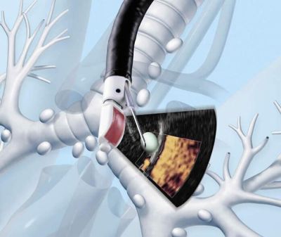
Endobronchial ultrasound (EBUS) is a bronchoscopy method that uses ultrasound to imagine structures close to the central airways (linear EBUS) and lung parenchyma (radial probe EBUS). EBUS is a minimally invasive but highly effective procedure used to diagnose lung cancer, infections, and other diseases, causing enlarged lymph nodes in the chest. It allows doctors to perform a method known as transbronchial needle aspiration (TBNA) to get a tissue or fluid samples from the lungs and surrounding lymph nodes without conventional surgery. Samples are used to diagnose and stage lung cancer, detect infections, and identify inflammatory diseases that affect the lungs, such as sarcoidosis or cancers like lymphoma.

When does a patient need an EBUS?

The main reason is that the doctor identified something unusual on an X-ray or other kind of test, or not feeling well for some time.

Requirements before and after EBUS:

You may need to stop taking some anticoagulants a few days before the test, but it is best to ask your doctor. Eat a light dinner the night before and NEVER eat or drink anything on the morning of the test. You will also need to remove any dentures, hearing aids, contact lenses, or eyeglasses you may be wearing. At the end of the test, you will eat or drink three hours later. It is also essential to be accompanied by a relative or a friend, since it is not advisable to drive after the examination.

Sleep Study

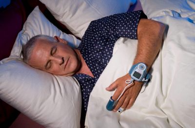
A Sleep Study is used to analyze Sleep Apnea, a sleep disorder that individuals have during their sleep. It is an overnight “in-home” sleep study in the comfort of your own bedroom while wearing a small portable device. Sleep Apnea is the sleep disorder that people face during sleep, which pauses in breathing or when low breathing occurs more often than usual. Every pause or stop of breathing can last from a few seconds up to a few minutes, and they might happen several times a night. This disorder, commonly known as snoring, and those usually affected, experience sleepiness, feel tired during the day, or even are hyperactive.

There are two (2) forms of Sleep Apnea, either obstructive sleep apnea (OSA), where a blockage of airflow disrupts breathing, or central sleep apnea (CSA) where frequent unconscious breath stops or might be a combination of both. The most common is obstructive sleep apnea.

The main reasons people suffer from sleep apnea, and most of the time are unaware of it, are overweight, allergies, family history, having a narrow upper airway, ineffective pharyngeal dilator muscle during sleep, unstable control of breathing, or airway narrowing during sleep.

The best way to treat sleep apnea is through vital lifestyle changes; stop alcohol and smoking, lose weight, use a breathing device, or even through surgery. If patients ignore or don’t pay any attention to this sleep disorder, they might face high risks of heart attack, stroke, diabetes, heart failure, irregular heartbeat, etc.

People suffering from sleep apnea in midlife have more chances of developing Alzheimer’s in older age. Also, sleep apnea is more common in men than women.
When does a patient need a sleep apnea test?

To identify the reasons for the breathing disorders during the night, as this might become vital for life.

Requirement before sleep apnea:

Book an appointment with your pulmonologist, requesting the sleep apnea test.

Spirometry

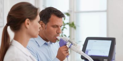
Spirometry is a commonly used test, performed by pulmonologists to assess how well lungs work by measuring how much air you inhale and exhale and how quickly you exhale. This test is used to diagnose asthma, chronic obstructive pulmonary disease, and other conditions that affect breathing.

The equipment that pulmonologists use for this test, called a spirometer, measures the volume of air inspired and expired by the lungs. A spirometer measures ventilation, the movement of air into and out of the lungs.

A spirometry results pneumatograph called a spirogram, which identifies two different abnormal ventilation patterns, obstructive and restrictive.

When does a patient need a spirometry test?

A patient needs to have spirometry to diagnose or manage asthma, detect lung illness when having breathlessness symptoms, distinguish respiratory from cardiac disease as the cause, and measure bronchial responsiveness in patients suspected of having asthma

Requirement before spirometry:

Book an appointment with your pulmonologist, requesting the spirometry test.

Cardiogram (ECG or EKG)

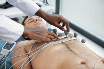
ECG or electrocardiogram, as most commonly known, records the electrical activity of the heart at rest by attaching electrodes to the surface of the skin of the patient, through a machine called ECG or Electrocardiograph. Cardiologists are using this test in order to interpret the electrical signals, which are triggered by an electrical impulse, as they travel through the heart. As these signals are recorded, doctors can check the patterns between heartbeats and rhythms to diagnose various heart conditions. ECG is being used to measure the rate and regularity of heartbeats, the position and size of the chambers, any damage on the heart, and the effects of drugs or devices used to control the heart, such as artificial pacemakers. Finally, it is a simple and painless test and the results of it can be reported on the same day, thus patients know immediately their results.

When does a patient need a cardiogram (ECG)?

An ECG estimates how the heart is functioning and it is wise to be performed to detect and prevent possible narrowing of the coronary arteries, a heart attack, or an irregular heartbeat like atrial fibrillation. What an ECG actually does is record how often the heart beats (heart rate) and how regularly it beats (heart rhythm).

Requirement before cardiogram (ECG):

Book an appointment with your cardiologist, requesting the cardiogram (ECG) test.

Cardiac ultrasound

Cardiac ultrasound, echocardiography, echocardiogram, or echo, is the heart’s ultrasound and is one of the best preventative examinations for the detection of cardiomyopathies. This test enables cardiologists to diagnose and treat suspected heart diseases, to view the size and shape of the heart, pumping capacity, diastolic function, and other helpful information.

In addition, stress echo may also indicate whether any chest pain or other related symptoms are associated with heart disease. The major advantage of echo is that it has no known risks or side effects and is not invasive.

Also, using a Doppler ultrasound, the doctor can accurately assess the blood flowing through the heart, and this allows doctors to check if the blood flow is normal or abnormal. With a color Doppler, Doctors can identify possible abnormal communications between the left and right sides of the heart, leaking of blood through the valves, as well as have a precise evaluation of the severity of aortic valve stenosis.

When does a patient need a cardiac ultrasound?

If having symptoms such as shortness of breath or chest pain, the doctors will have to examine for potential problems with the valves or chambers of the heart.

Requirement before cardiac ultrasound:

Book an appointment with your cardiologist, requesting the cardiac ultrasound test.

Stress Test

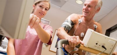
A stress test is a test cardiologists use to estimate a patient’s potential risks of having heart disease. It is a simple test since individuals walk on a treadmill or pedal on a static bike, and as you go along, it gets more difficult. Through the stress test, the patient’s electrocardiogram, heart rate, and blood pressure are being tracked. Therefore, the doctor can check how much a patient’s heart can operate before an abnormal rhythm starts of blood flow to the heart muscle drops; in conclusion, doctors can identify how the heart responds when stressed.

Although there are various stress tests, the most commonly used is the treadmill test, where doctors can check how the heart responds while being pushed.

When does a patient need a stress test?

Doctors can assess symptoms like chest pain, shortness of breath, examine if enough blood flows in the heart, identify possible abnormal heart rhythms or coronary heart disease, and, in case the patient receives medication, check if it is working.

Requirement before stress test:

Patients should not eat or drink anything for at least 4 hours before the test. It is also wise not to have anything that contains caffeine and not to take (in case patients receive any) heart medication on the day of the test unless the doctors advise you opposite.

Bone Density Measurement?

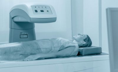
Bone Density Measurement or DΕXA (Dual-Energy X-Ray Absorptiometry) is the test that uses X-rays to determine bone mass. Considered the best test to check bone density, it is widely used to diagnose osteoporosis.

Osteoporosis is a chronic disease of bone metabolism slowly developing over several years. There is a gradual decrease in density and quality of the bone in women AND men, resulting in bones becoming more brittle and thinner. Based on the above, there is a greater risk of bone fracture as their strength and elasticity decrease. Osteoporosis is preventable in most cases.

Prevention is everything; for this reason, women after menopause and men after a certain age, are right to perform this test, regardless of having bone or spine problems.

When does a patient need a bone density measurement?

The main reason for having the test is to find bone loss, follow treatment and prevent fractures and disability.

Requirement before a bone density measurement:

Book an appointment with your doctor, requesting the bone density measurement test. It is also wise to avoid taking calcium supplements at least one day before the test.

X-ray Department

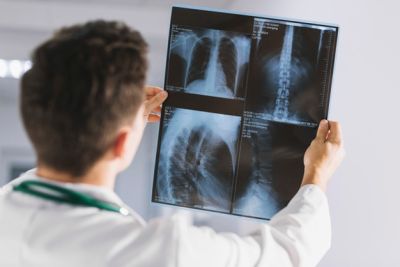
An x-ray examination creates images of your internal organs or bones to help diagnose conditions or injuries. A special machine emits (puts out) a small amount of ionising radiation. This radiation passes through your body and is captured on a special device to produce the image.The X-Ray Department of EIMC Clinics is fully equipped with state-of-the-art radiological equipment as well as highly trained staff.

Contact Us

Please contact us for more information and to schedule a meeting

Please enable JavaScript in your browser to complete this form.
contact form

Opening hours

Mon-Fri 08:30 – 18:00 / Sat-Sun closed

© EIMC Clinic: Leading Medical Center In Nicosia. All rights reserved.